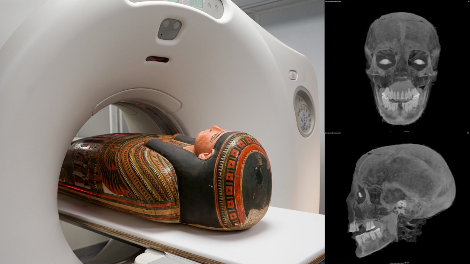
The Chicago Field Museum is home to more than a dozen ancient Egyptian mummies, but one in particular has baffled researchers for years. Now the mystery of Lady Chenet-aa’s funeral proceedings appears to have been solved with the help of a CT scanner.
Lady Chenet-aa lived about 3,000 years ago during the 22nd Dynasty during Egypt’s Third Intermediate Period. Shortly after her death, one of the ways funeral experts prepared her for the afterlife was to build a cartonnage: a papier-mâché-like box that held the body of a deceased person. In the case of Chenet-aa, however, there is no visible seam, leading Egyptologists to wonder for years how exactly embalmers placed her in the casing. According to one Announcement October 24 from the Field Museum, a mobile CT scanner finally helped explain the strategy behind Chenet-aa’s “locked mummy” cartonnage, as well as new physical information about her at the time of her death.

Computed tomography or CT scanning creates 3D views of a subject by digitally stacking thousands of X-ray scans. Over four days, researchers recently transported 26 mummies (including Chenet-aa) to a mobile machine parked outside the Field Museum. The resulting images allowed experts to analyze the cartonnage and its contents at an unprecedented level of detail, helping them learn how funeral directors placed Chenet-aa in what appeared to be a seamless casing in the first place.
“You can start to see that there is a seam running down the back and some laces,” JP Brown, the museum’s senior curator of anthropology, said in a statement.

The CT images also revealed new details about Chenet-aa’s health during her final days. According to investigators, the aristocrat died in his late 30s or early 40s, although the cause of death was not specified. Her teeth weren’t in the best condition; many were missing at the time of death, while the remaining teeth showed “significant wear,” the museum’s announcement said. Experts attribute this to a diet that often included ‘stray grains of sand’ in foods that were harmful to tooth enamel.

Images of the mummy’s skull include bright objects in both eye sockets, despite an apparent lack of organs. These are artificial eyes of unknown material, intended to enable Chenet-aa vision in the afterlife.
[Related: ‘Screaming woman’ may solve a 3,500-year-old mummy mystery.]
“The ancient Egyptian view of the afterlife corresponds with our ideas about retirement savings. It’s something you prepare for, put money aside all your life and hope that in the end you have enough to really enjoy,” Brown explained. “The additions are very literal. If you want eyes, there must be physical eyes, or at least a physical allusion to eyes.”
These are just the first of many possible insights into some of the Field Museum’s oldest and most delicate remains. Over the next year, researchers hope to continue examining the thousands of CT scans for more clues about death – and life – in ancient Egypt.


Leave a Reply